The TMJ examination conists of several routine exams. These exams help us to evaluate the condition of the temporomandibular joint and the surrounding structures and to help determine the best solutions moving forward.
The TMJ examination begins with palpation of the muscles in the face and neck regions. These regions include the elevator muscles, depressor muscles, cervical muscles and intraoral muscles. If the patient has a history of fibromyalgia, pain points around the rest of the body may be examined. Patients with more acute temporomandibular joint disorders have the majority of muscle pain in the elevator muscles.
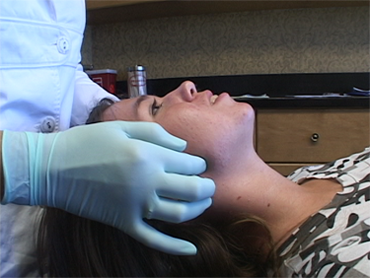
The TMJ examination begins with palpating the muscles in the face and neck.
This exam includes an assessment of the prematurities, slides and interferences present in the bite.
In this exam, the oral soft tissues are checked for lesions and masses; the cervical region is checked for lymphadenopathy, thyromegaly and vascular disease; the salivary glands are palpated for enlargement, tenderness and masses; the facial skeleton is examined for distortions; the midlines of the nasion, the nasal tip, philtrum of the upper lip and chin are marked; the palpated angle of the mandible is marked; facial measurements are recorded; the distance from the medial canthus of the orbit to the incisal edge of the maxillary cuspid on each side is measursed using a Boley guage; and the ramus height is measured from the top of the tragus to the angle on side of the mandible. These measurements allow us to assess the face for occlusal plane canting, ramus asymmetry, and midline discrepancies of the skeleton and the maxillary and mandibular teeth. These measurements give a good assessment of potential fossa ramus deficiencies from a disordered temporomandibular joint.
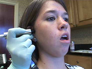
Doppler Auscultation is used to listen to the sounds in the joint.
We use Doppler Auscultation to determine the alignment of the disk inside the temporomandibular joint by listening to the movements of the structures inside.
Range of Motion Measurements - We use a Therabite Ruler for measuring the range of motion. We ask patients to open their mouth as far as possible, with or without pain, for the full opening.
This exam includes the assessment of unmanipulated and manipulated prematurities. We record the degree of overjet, overbite, open bite and cross bite. The midlines, Angle Classification and Dawson Classification are also recorded.
The examination of the temporomandibular joint is performed through palpation, bimanual manipulation, and load testing. The lateral part of the condyle is palpated for tenderness and capsulitis; from the ear canal, the posterior part of the joint can be assessed for tenderness associated with retrodiskitis and for laxity of the lateral collateral ligament. Palpable clicking is noted for each joint. The last part of the exam includes load testing through bimanual manipulation. This part of the exam is used to verify the Dawson Classification.
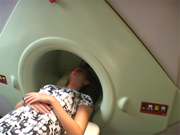
Diagnostic Image includes CT and Magnetic Resonance scans.
We use radiographs (Panorex, Transcranials, Tomographs and CTs) to assess the temporomandibular joint. Panorex radiographs serve as an overall screening examination for hard and soft tissue lesion of the facial skeleton. Transcranials radiographs are excellent diagnostic tools for screening join space alterations and arthritic patterns in the temporomandibular joint. Tomography gives a better assessment of joint spacing and joint surface contours. CT scanning allows for assessment of the temporomandibular join from any angle or plane. We use a 3D CT scanner, which makes it much easier for the patient and practioner to visualize all imaged structures.
MRI scanning is similar to CT scanning in that a 3D image of the patient’s anatomy is produced for assessment. However, MRI scans allow the doctor to assess soft tissue and liquid that is not visible in CT scans. MRIs are particularly necessary during the assessment of the TMJ patient. They are able to show the condition of the TMJ disk and surrounding anatomy, allowing the doctor to provide the patient with the most accurate information to make an informed decision.
727-823-3220
131 2nd Ave. S.
St. Petersburg, FL 33701